
Early diagnosis shapes outcomes for dogs facing cancer concerns. This article explains how veterinarians identify tumors, what each diagnostic step means, and why timely testing matters. You will learn how exams, imaging, lab work, and biopsies work together to confirm disease and guide care.
Neoplasia in dogs appears in many forms, so clear evaluation protects comfort, safety, and quality of life.
Understanding Neoplasia and Why Diagnosis Matters
Neoplasia refers to abnormal cell growth that forms a mass or spreads into nearby tissue. Some growths remain slow and harmless. Others invade organs or spread through blood and lymph. Diagnosis determines whether a lump poses a risk and how urgent care becomes. Clear answers reduce stress and prevent delays.
Veterinarians follow a structured process that balances speed and accuracy. Each step adds facts. Skipping steps risks false assumptions. A careful plan leads to decisions that match the dog, the family, and the condition.
The Initial Veterinary Exam
A thorough exam starts the process. Veterinarians review history, lifestyle, and recent changes. Appetite shifts, weight loss, pain, coughing, or skin changes guide next steps. Hands-on checks reveal size, firmness, heat, and mobility of masses. Lymph nodes receive close attention.
This visit sets priorities. Some findings call for monitoring. Others demand rapid testing. The exam also helps rule out infection or injury that can mimic cancer.
Baseline Testing and Lab Work
Blood and urine tests create a health snapshot. They reveal anemia, infection, organ stress, and clotting risks. Results influence anesthesia safety and imaging choices. Baseline values help track change over time.
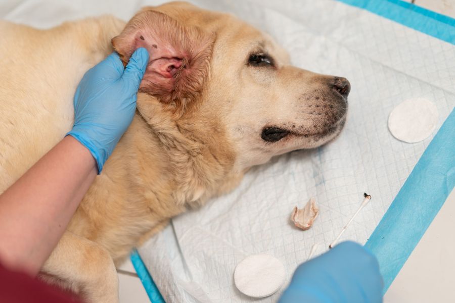
Cytology as a First Look
Fine needle aspiration collects cells using a small needle. The method causes minimal discomfort and rarely needs sedation. A pathologist reviews cell shape and patterns to classify growths as benign, malignant, or unclear.
Cytology answers many questions quickly. Some tumors shed clear cells that allow a confident diagnosis. Others require deeper sampling. A non-diagnostic result still informs the plan and avoids delays.
Imaging to See the Full Picture
Imaging shows size, location, and spread. Radiographs assess the lungs and bones. Ultrasound evaluates organs and guides sampling. Advanced imaging, such as CT or MRI, maps complex areas like the head or chest.
These tools support staging. Staging describes how far the disease extends. Accurate staging shapes treatment options and expectations. Imaging also helps surgeons plan safer procedures.
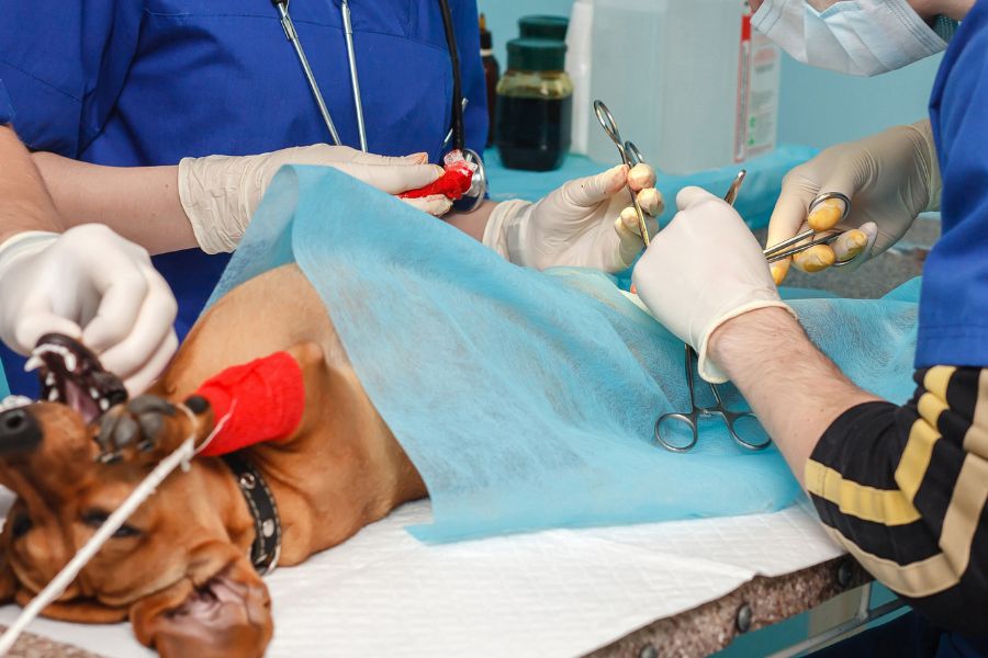
Biopsy for Definitive Diagnosis
A biopsy removes tissue for detailed review. This step confirms tumor type and grade. Grade reflects how aggressive cells appear under the microscope. Incisional biopsies sample part of a mass. Excisional biopsies remove the entire growth when appropriate.
Biopsy results guide next steps with clarity. They support choices about surgery, medication, radiation, or comfort care. For many families, this report brings relief through certainty.
When Referral Makes Sense
Complex cases benefit from specialists. Oncologists, surgeons, and internists add experience with rare tumors and advanced tools. Referral supports dogs with tumors near vital structures or with signs of spread.
Staging and Treatment Planning
Staging combines exam findings, lab work, imaging, and biopsy results. The care team outlines options with clear goals. Some plans aim to cure. Others control disease or relieve pain. Honest discussion supports informed consent and shared decisions.
Throughout planning, quality of life stays central. Dogs live in the present. Treatments balance benefit with comfort. Monitoring plans include rechecks and imaging as needed.
Common Questions About Diagnostic Safety
Most diagnostic steps carry low risk. Fine needle aspiration rarely causes bleeding. Imaging avoids excess exposure. Biopsies use pain control and careful technique. The team explains risks and benefits before each step.
Neoplasia in dogs requires thoughtful evaluation rather than assumptions. A stepwise approach protects dogs and supports families with clear answers.
Working With Your Veterinary Team
Open communication supports accurate diagnosis. Share timelines, photos of changes, and prior records. Ask direct questions about goals, costs, and follow-up. Keep a simple log of appetite, energy, and pain signs at home. These details help refine decisions.
Consistency matters after testing. Attend rechecks on schedule. Repeat imaging or lab work when advised. Small changes over time can signal response or progression. Early notice allows timely adjustments.
Emotional support also matters. Families face worry while waiting for results. Veterinary teams explain findings in plain language and respect values. Clear plans reduce uncertainty and help everyone move forward with confidence and care.
Taking the Next Step
Trust the process and take notes during visits. Written summaries help recall options and instructions later. Reliable diagnosis depends on teamwork, patience, and thoughtful follow-through that keeps the dog comfortable while decisions unfold.
Your dog deserves clarity and comfort at every step.
If you notice a lump, behavior change, or unexplained symptom, timely evaluation matters. Clear diagnosis guides calm decisions and protects quality of life. Neoplasia in dogs benefits from early, structured assessment. Book an appointment with our North Charleston vets to discuss concerns and plan appropriate testing today with confidence.
Frequently Asked Questions(FAQs):
1. How soon should a new lump be checked?
A: Any new lump deserves an exam within weeks. Rapid growth, pain, bleeding, or illness requires prompt visits. Early checks improve options and reduce anxiety while establishing a clear baseline for monitoring.
2. Does every lump need a biopsy?
A: No. Many lumps receive a diagnosis through exam and cytology. Biopsy becomes important when results remain unclear or when treatment decisions depend on tumor type and grade.
3. Is sedation always needed for biopsies?
A: Sedation depends on location and size. Small skin biopsies may need local anesthesia. Deeper samples often require sedation to ensure comfort and accuracy.
4. Can imaging replace a biopsy?
A: Imaging shows structure and spread, but cannot confirm cell type. A biopsy provides a definitive diagnosis. Both tools work together to guide accurate care.
5. How long do results take?
A: Cytology results often return within days. Biopsy reports may take one to two weeks. Timelines vary by lab and complexity.
6. What if the results are inconclusive?
A: Inconclusive findings lead to repeat sampling or advanced imaging. Clear communication helps families understand next steps without panic or delay.